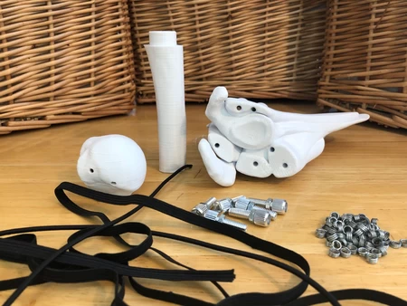
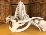
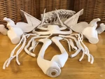
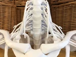
16-piece full size brachial plexus model 3D for print

7314 Views 1 Likes 0 Downloads Download the piece here from 3dforprint
S...ee here for video: https://youtu.be/Uol33HA0QOc
See here for stable download: https://thangs.com/mythangs/file/47926
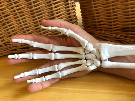
The brachial plexus is a network of nerves in the shoulder that carries movement and sensory signals from the spinal cord to the arms and hands. This model is designed for use as a clinical education tool, specifically for ultrasound guided nerve blocks and anesthesia for surgery.
The model can be used upright or in supine. You can print either shoulder or both. Use different coloured filament to emphasis structures.
The “Brachial Plexus” includes all roots, trunks, divisions, cords and branches, as well as proximal portions of the suprascapular and long thoracic nerve. The phrenic nerve is included as well, not because it is a part of the brachial plexus, but because it is a structure to identify and avoid when doing nerve blocks or surgery in this area.
The main structural pieces are attached with magnets. To assemble both sides with magnets, you will need 36 1/4” x 1/8” (6mm x 3mm) cylindrical neodymium magnets. www.kjmagnetics.com is a good source of magnets. Pieces can be glued together for permanent attachment.
The roots of the brachial plexus and phrenic nerve are attached via insertion holes in the intervertebral foramen. The brachial plexus and subclavian/axillary artery are also held in place by the clavicle.
The following pieces form the structural base:
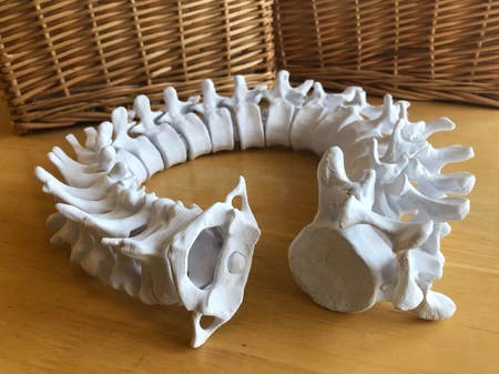
• Spine Base
• Right and/or Left Scapula Rhomboid Base
• Sternum Rib Base
• Right and/or Left Clavicle
The follow pieces are non-structural. Print the pieces and sides that you need.:
• Head of Humerus
• Anterior Scalene
• Brachial Plexus
• Phrenic Nerve
• Subclavian/Axillary Artery
This is an advanced print requiring knowledge in setting up supports and post-print cleanup.
The “Spine Base” and “Scapula Rhomboid Bases” should be printed as provided. The remaining files are in a “suggested orientation,” you may need to reorient and prepare them according to your preferences, equipment, and skill level. In general, try to orient outer surfaces upwards so that the surfaces in contact with support structures are less visible in the final model.
For pieces with contoured bottom surfaces and hard-to-reach support areas, try raising the model a few millimeters off the build plate and using “tree supports.”
Here are considerations for assembly:
• Before inserting magnets, clean out the magnet anchors with a 1/4” drill bit to remove any imperfections, allowing the magnets to sit properly.
• Ensure that you insert magnets with polarity oriented to allow proper connections.
• You may need to use glue to secure some of the magnets in place, especially if using the smaller metric sizes. CA glue works well.
• Expect to do some post-print cleanup with sandpaper and files.
Every attempt has been made to maintain anatomical correctness; however, the following small modifications were necessary to optimize the files for 3D printing:
• Magnet anchors have been added to allow insertion of magnets.
• Some areas of bone have been thickened to support the magnet anchors and add strength
• A portion of the posterior spine of the scapula has been enlarged to support the model in supine.
• All neural structures have been thickened for strength and to facilitate printing.
• The brachial plexus has been reshaped in some areas to allow for ease of printing and assembly. These alterations are within the range of normal anatomical variation.
• Anterior scalene is positioned slightly anterior to create more space in the interscalene triangle.
• Groves have been added on the first ribs to facilitate proper positioning of the subclavian artery.
This project was done in collaboration with Eric Kramer and colleagues.
Eric Kramer is a nurse anesthesiologist and nurse practitioner with an interest in point of care ultrasound and ultrasound guided regional anesthesia. He is faculty for National Universities doctoral nurse anesthesiology program and is also the founder of Open Source Medicine (learn.osmedicine.org), a nonprofit geared towards training national anesthetists in developing countries.
This project uses reference files from BodyParts3D, © The Database Center for Life Science licensed under CC Attribution-Share Alike 2.1 Japan
Designer
DaveMakesStuff3d model description
This 16-piece, full-sized anatomical model shows the position of the brachial plexus nerves relative to surrounding bones, muscles, and arteries.S...ee here for video: https://youtu.be/Uol33HA0QOc
See here for stable download: https://thangs.com/mythangs/file/47926
The brachial plexus is a network of nerves in the shoulder that carries movement and sensory signals from the spinal cord to the arms and hands. This model is designed for use as a clinical education tool, specifically for ultrasound guided nerve blocks and anesthesia for surgery.
The model can be used upright or in supine. You can print either shoulder or both. Use different coloured filament to emphasis structures.
The “Brachial Plexus” includes all roots, trunks, divisions, cords and branches, as well as proximal portions of the suprascapular and long thoracic nerve. The phrenic nerve is included as well, not because it is a part of the brachial plexus, but because it is a structure to identify and avoid when doing nerve blocks or surgery in this area.
The main structural pieces are attached with magnets. To assemble both sides with magnets, you will need 36 1/4” x 1/8” (6mm x 3mm) cylindrical neodymium magnets. www.kjmagnetics.com is a good source of magnets. Pieces can be glued together for permanent attachment.
The roots of the brachial plexus and phrenic nerve are attached via insertion holes in the intervertebral foramen. The brachial plexus and subclavian/axillary artery are also held in place by the clavicle.
The following pieces form the structural base:
• Spine Base
• Right and/or Left Scapula Rhomboid Base
• Sternum Rib Base
• Right and/or Left Clavicle
The follow pieces are non-structural. Print the pieces and sides that you need.:
• Head of Humerus
• Anterior Scalene
• Brachial Plexus
• Phrenic Nerve
• Subclavian/Axillary Artery
This is an advanced print requiring knowledge in setting up supports and post-print cleanup.
The “Spine Base” and “Scapula Rhomboid Bases” should be printed as provided. The remaining files are in a “suggested orientation,” you may need to reorient and prepare them according to your preferences, equipment, and skill level. In general, try to orient outer surfaces upwards so that the surfaces in contact with support structures are less visible in the final model.
For pieces with contoured bottom surfaces and hard-to-reach support areas, try raising the model a few millimeters off the build plate and using “tree supports.”
Here are considerations for assembly:
• Before inserting magnets, clean out the magnet anchors with a 1/4” drill bit to remove any imperfections, allowing the magnets to sit properly.
• Ensure that you insert magnets with polarity oriented to allow proper connections.
• You may need to use glue to secure some of the magnets in place, especially if using the smaller metric sizes. CA glue works well.
• Expect to do some post-print cleanup with sandpaper and files.
Every attempt has been made to maintain anatomical correctness; however, the following small modifications were necessary to optimize the files for 3D printing:
• Magnet anchors have been added to allow insertion of magnets.
• Some areas of bone have been thickened to support the magnet anchors and add strength
• A portion of the posterior spine of the scapula has been enlarged to support the model in supine.
• All neural structures have been thickened for strength and to facilitate printing.
• The brachial plexus has been reshaped in some areas to allow for ease of printing and assembly. These alterations are within the range of normal anatomical variation.
• Anterior scalene is positioned slightly anterior to create more space in the interscalene triangle.
• Groves have been added on the first ribs to facilitate proper positioning of the subclavian artery.
This project was done in collaboration with Eric Kramer and colleagues.
Eric Kramer is a nurse anesthesiologist and nurse practitioner with an interest in point of care ultrasound and ultrasound guided regional anesthesia. He is faculty for National Universities doctoral nurse anesthesiology program and is also the founder of Open Source Medicine (learn.osmedicine.org), a nonprofit geared towards training national anesthetists in developing countries.
This project uses reference files from BodyParts3D, © The Database Center for Life Science licensed under CC Attribution-Share Alike 2.1 Japan